Home
Uncategories
Long Bone Labeled / Anatomy Gross Anatomy Physiology Cells Cytology Cell Physiology Organelles Tissues Histology Organs Regional Anatomy Organ / Long bones of the leg include the femur, tibia, fibula, metatarsals, and phalanges.
Long Bone Labeled / Anatomy Gross Anatomy Physiology Cells Cytology Cell Physiology Organelles Tissues Histology Organs Regional Anatomy Organ / Long bones of the leg include the femur, tibia, fibula, metatarsals, and phalanges.
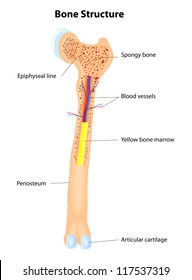
Long Bone Labeled / Anatomy Gross Anatomy Physiology Cells Cytology Cell Physiology Organelles Tissues Histology Organs Regional Anatomy Organ / Long bones of the leg include the femur, tibia, fibula, metatarsals, and phalanges.. While the name suggests a larger size of bone, bones such as the metacarpals in the fingers are classified as long bones. Long, short, flat, irregular and sesamoid. The walls of the diaphysis are composed of dense and hard compact bone. Red bone marrow fills the spaces between the spongy bone in some long bones. The structure of a long bone:
The bones typically consist of a long shaft called the diaphysis, and two wider extremities on the ends called epiphyses. The diaphysis is the tubular shaft that runs between the proximal and distal ends of the bone. They are one of five types of bones: Human anatomy for muscle, reproductive, and skeleton. The long bones have a long, central shaft that enlarges at the ends into epiphysis.
Long Bone Anatomy Images Stock Photos Vectors Shutterstock from image.shutterstock.com Related posts of long bone label chart human back muscles and bones. The long bones in the legs are the femur, tibia, and fibula. Learn vocabulary, terms, and more with flashcards, games, and other study tools. The long bones have a long, central shaft that enlarges at the ends into epiphysis. Long bones of the leg include the femur, tibia, fibula, metatarsals, and phalanges. Anatomical diagram of internal organs. The bones of the hands can be divided into those that make up the upper arm, the lower arm, the wrist, the palm and the fingers. The bones typically consist of a long shaft called the diaphysis, and two wider extremities on the ends called epiphyses.
New users enjoy 60% off.
All of the bones in the arms and legs, except the patella, and bones of the wrist, and ankle, are long bones. The long bones in the legs are the femur, tibia, and fibula. Label the parts of a long bone. Keep in mind, however, that the term describes the shape of a bone, not its size. A long bone is one that is cylindrical in shape, being longer than it is wide. The bones typically consist of a long shaft called the diaphysis, and two wider extremities on the ends called epiphyses. The long bones (ossa longa) are those that are longer than they are wide. A long bone has two parts: A typical long bone showing gross anatomical features. Labelled image long bones are the most common bones found in the human body. Bone on side of the foot 12 photos of the bone on side of the foot bone on side of foot growing, bone. The wider section at each end of the bone is called the epiphysis (plural = epiphyses), which is filled internally with spongy bone, another type of osseous tissue. This is due to the shape of the bones, not their size.
Labelled image long bones are the most common bones found in the human body. The clavicles (collar bones) are also long bones. The bones of the hands can be divided into those that make up the upper arm, the lower arm, the wrist, the palm and the fingers. Label the parts of a long bone. The long bones (ossa longa) are those that are longer than they are wide.
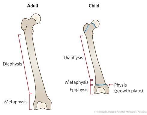
Fracture Education Anatomic Differences Child Vs Adult from www.rch.org.au This is an online quiz called label a long bone there is a printable worksheet available for download here so you can take the quiz with pen and paper. Anatomical diagram of internal organs. The long bones have a long, central shaft that enlarges at the ends into epiphysis. The structure of a long bone allows for the best visualization of all of the parts of a bone (figure 1). Labelled diagram of long bone. It runs from the shoulder to the elbow. Long bones of the leg include the femur, tibia, fibula, metatarsals, and phalanges. All of the bones in the arms and legs, except the patella, and bones of the wrist, and ankle, are long bones.
Anatomical diagram of internal organs.
Bone · august 3, 2016. These bones develop via endochondral ossification, a process in which the hyaline cartilage plate is slowly replaced.a shaft, or diaphysis, connects the two ends known as the epiphyses (plural for epiphysis). Labelled diagram of long bone. The diaphysis and the epiphysis. Label the parts of a long bone. Long bones include the humerus (upper arm), radius (forearm), ulna (forearm), femur (thigh), fibula (thin bone of the lower leg), tibia (shin bone), phalanges (digital bones in the hands and feet), metacarpals (long bones within the hand), and metatarsals (long bones within the feet). Related posts of long bone diagram labeled muscles and bones of the chest. The long bones (ossa longa) are those that are longer than they are wide. A long bone has two parts: Download 41 long bone labeled stock illustrations, vectors & clipart for free or amazingly low rates! Related posts of labeled diagram of long bone bone in arm pictures. Anatomical diagram of internal organs. The long bones in the legs are the femur, tibia, and fibula.
Download 41 long bone labeled stock illustrations, vectors & clipart for free or amazingly low rates! A long bone has two parts: The bones of the hands can be divided into those that make up the upper arm, the lower arm, the wrist, the palm and the fingers. Bone on side of the foot 12 photos of the bone on side of the foot bone on side of foot growing, bone. New users enjoy 60% off.
Where Is The Proximal Epiphysis Located from o.quizlet.com A typical long bone shows the gross anatomical characteristics of bone. It is very strong to support the body's weight, made up mostly of compact bone and some inner spongy bone (described below). Humerus (2) radius (2) ulna (2) carpals (16) metacarpals (10) phalanges (28) total number of bones=60. Long bones of the leg include the femur, tibia, fibula, metatarsals, and phalanges. The bones of the hands can be divided into those that make up the upper arm, the lower arm, the wrist, the palm and the fingers. Labelled image long bones are the most common bones found in the human body. Types of bone long bones. Labelled diagram of long bone.
Bone in arm pictures 12 photos of the bone in arm pictures bone cancer arm pictures, pictures of bone cancer in arm, bone, bone cancer arm pictures, pictures of bone cancer in arm
A long bone is one that is cylindrical in shape, being longer than it is wide. Labelled diagram of long bone. G = medullary cavity (yellow marrow) h = endosteum. The bones typically consist of a long shaft called the diaphysis, and two wider extremities on the ends called epiphyses. Muscles and bones of the chest 12 photos of the muscles and bones of the chest muscles and bones in chest, muscles and bones of the chest, muscular or bone problems of the chest wall, bone, muscles and bones in chest, muscles and bones of the chest, muscular or bone. New users enjoy 60% off. Long bones include the humerus (upper arm), radius (forearm), ulna (forearm), femur (thigh), fibula (thin bone of the lower leg), tibia (shin bone), phalanges (digital bones in the hands and feet), metacarpals (long bones within the hand), and metatarsals (long bones within the feet). Related posts of long bone diagram labeled muscles and bones of the chest. The walls of the diaphysis are composed of dense and hard compact bone. Related posts of labeled diagram of long bone bone in arm pictures. This is a single long bone of the upper arm. They are composed mostly of compact bone, and are roughly cylindrical in shape with enlarged ends filled with spongy bone. Long, short, flat, irregular and sesamoid.

0 Comments:
Posting Komentar