Home
Uncategories
Back Of Neck Anatomy Bones : Three Keys to Solving Chronic Neck Pain | Impulse Sport Therapeutics - Facet joints connect each vertebra, with fluid supporting.
Back Of Neck Anatomy Bones : Three Keys to Solving Chronic Neck Pain | Impulse Sport Therapeutics - Facet joints connect each vertebra, with fluid supporting.
Back Of Neck Anatomy Bones : Three Keys to Solving Chronic Neck Pain | Impulse Sport Therapeutics - Facet joints connect each vertebra, with fluid supporting.. Bones of the chest and upper back from www.innerbody.com this article describes the anatomy of the head and neck of the human body, including the brain, bones, muscles, blood vessels, nerves, glands, nose, mouth, teeth, tongue, and throat. The anterior, and the posterior, triangles of the neck. An area called the occiput. Go ahead, see if you can find it. The neck contains seven of.
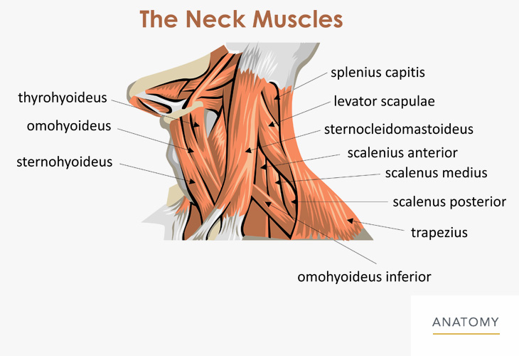
Go ahead, see if you can find it. Then it extends to the clavicles and the sternum in front. In the back, the neck reaches the c7 vertebra. The anterior triangle of the neck is made by the anterior border of the sternocleidomastoid muscle, the inferior border of the mandible and the midline of the neck. The majority of neck muscles attach to the hyoid, separating them into two groups:
Bones of the Chest and Upper Back from www.innerbody.com From the sides and the back of the neck, the splenius. The neck is the part of the body that acts as a bridge between the trunk and the head. An area called the occiput. Browse 3,081 anatomy of neck and shoulder stock photos and images available, or start a new search to explore more stock photos and images. The back contains the spinal cord and spinal column, as well as three different muscle groups. Surface anatomy of the head and neck. Level i cervical lymph nodes. C3 to c6 are the typical cervical vertebrae characterised by the presence of transverse foramina and, in many people, by their bifid spinous processes.
The majority of neck muscles attach to the hyoid, separating them into two groups:
The 5 anatomical spaces of the infrahyoid neck. The bones of the neck include the seven cervical vertebrae, the hyoid bone, the clavicles and the manubrium of the sternum. From the sides and the back of the neck, the splenius. Level i cervical lymph nodes. The suprahyoid and infrahyoid muscles. There are two main triangles; The top of the cervical spine connects to the skull, and the bottom connects to the upper back at about shoulder level. The neck is the start of the spinal column and spinal cord. Long bones function to support the weight of the body and facilitate movement. The bone that rests on top of your spine. The skull can be further subdivided into: Head neck spine back abdomen chest shoulder arm elbow forearm hand wrist lung. Go ahead, see if you can find it.
Each of these bones is stacked one on top of the other and in between them rest structures called discs. The bones are separated by discs, made up of concentric circles of fibrous material with a bubble of gel in the centre, acting as shock absorbers for the all bouncing, jogging, jumping movements of the neck. See easy levator scapulae stretch for neck pain Hyoid bone explore study unit .anatomy of the body neck anatomy hyoid bone neck anatomy landmarks anterior neck bones arthritis neck.
Cervical Spine Anatomy (Neck) from www.spineuniverse.com Go ahead, see if you can find it. Heart pelvis hip thigh leg. C3 to c6 are the typical cervical vertebrae characterised by the presence of transverse foramina and, in many people, by their bifid spinous processes. The spine is composed of 33 bones called vertebrae, which stack together to form the spinal canal. The anterior triangle of the neck is made by the anterior border of the sternocleidomastoid muscle, the inferior border of the mandible and the midline of the neck. Long bones function to support the weight of the body and facilitate movement. The submandibular gland duct, or whartons duct, ends in the floor of mouth and is typically blocked when cancer invades in this area. Bones of the chest and upper back from www.innerbody.com this article describes the anatomy of the head and neck of the human body, including the brain, bones, muscles, blood vessels, nerves, glands, nose, mouth, teeth, tongue, and throat.
The skeletal section of the head and neck forms the top part of the axial skeleton and is made up of the skull, hyoid bone, auditory ossicles, and cervical spine.
The cervical vertebrae's main job is to support your head. Head neck spine back abdomen chest shoulder arm elbow forearm hand wrist lung. However, the muscles of the neck can also be easily strained or injured. Bone comprises the structure of the skeletal system and provides lever arms for locomotion. Close up macro view of human skull bone showing the anatomy of nasal foramen, nasal septum and orbital cavity. The bones of the chest and upper back combine to form the strong, protective rib cage around the vital thoracic organs such as the heart and lungs. The cervical spine and the hyoid bone constitute the bones of the neck. The neck begins at the lower edge of the jaw and the occipital bone, which is the base of the skull. In suspected cases of departure, the evaluation of hyoid bone is of great medicolegal value, because fracture of hyoid bone in such cases indicates departure by throttling or strangulation. The bone that rests on top of your spine. The anterior triangle of the neck is made by the anterior border of the sternocleidomastoid muscle, the inferior border of the mandible and the midline of the neck. The occipital bone is the only bone in your head that connects with your cervical spine (neck). The rib cage also anchors the bones of the head, neck, shoulders, and arms to the trunk of the body.
The neck is connected to the upper back through a series of seven vertebral segments. The anterior triangle of the neck is made by the anterior border of the sternocleidomastoid muscle, the inferior border of the mandible and the midline of the neck. Facet joints connect each vertebra, with fluid supporting. The anchor point of the neck is the hyoid bone, which is situated at the level of the 'adam's apple' (laryngeal prominence) in males. This nodal level can be subdivided into 1a (submental) and 1b (submandibular)drains from the lips, gum, teeth, tongue, anterior hard palate.
Neck / Back - Orthopedic Specialist of Northern California - Orthopedic Specialist of Northern ... from www.oanc.org The cervical spine is comprised of the 7 uppermost vertebrae of the vertebral column. C3 to c6 are the typical cervical vertebrae characterised by the presence of transverse foramina and, in many people, by their bifid spinous processes. Go ahead, see if you can find it. Back of neck anatomy bones. The anchor point of the neck is the hyoid bone, which is situated at the level of the 'adam's apple' (laryngeal prominence) in males. The occipital bone is a bone that covers the back of your head; The skeletal section of the head and neck forms the top part of the axial skeleton and is made up of the skull, hyoid bone, auditory ossicles, and cervical spine. The bones of the head and neck play the vital role of supporting the brain, sensory organs, nerves, and blood vessels of the head and protecting these structures from mechanical damage.
Hyoid bone explore study unit
See easy levator scapulae stretch for neck pain The head rests on the top part of the vertebral column, with the skull joining at c1 (the first cervical vertebra known as the atlas). Level i cervical lymph nodes. However, the muscles of the neck can also be easily strained or injured. The bones of the neck serve to protect the spinal cord and to provide a site of attachment for the numerous neck muscles. The skeletal section of the head and neck forms the top part of the axial skeleton and is made up of the skull, hyoid bone, auditory ossicles, and cervical spine. Pain in a man's body pain in a man's body on a gray background. Facet joints connect each vertebra, with fluid supporting. The spine is made of 33 individual bones stacked one on top of the other. Close up macro view of human skull bone showing the anatomy of nasal foramen, nasal septum and orbital cavity. In the back, the neck reaches the c7 vertebra. An overview of the anatomy of the hand, including the bones of the hand, muscles, blood supply and nerve supply. Human bone is stretching arm and leg (whole body :
This protects the spinal cord inside back of neck anatomy. With the weight that must be supported, these muscles are strong.


0 Comments:
Posting Komentar